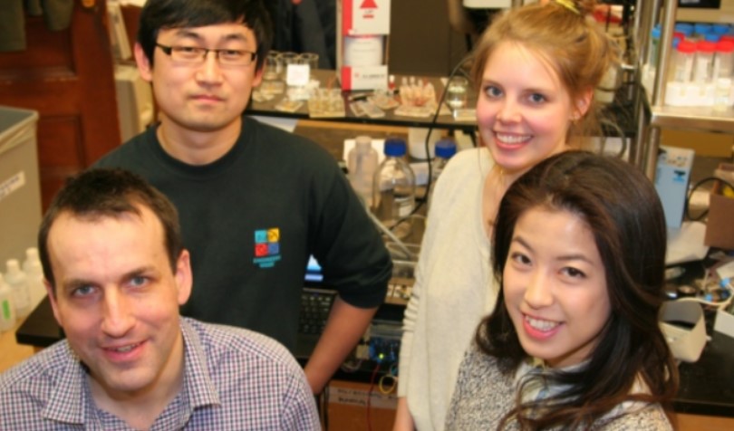
Dr. Marc Jeschke, the head of one of
Canada's largest burn treatment centers, had to admit the 3D skin
printer in his hands didn't look revolutionary."I actually find
it kind of fish-tanky," he told CBS News, laughing. But this boxy
prototype could change the way burns are treated, from current skin
grafting methods Jeschke calls "barbaric" to a process his team believes
will be faster, cheaper and easier on the patient, with an end result
-- functional human skin -- promising to be just like the real thing.
"It's cutting edge," said Jeschke, the director of Ross Tilley Burn Centre
at Sunnybrook Health Sciences Centre in Toronto, whose team developed
the process and printer in collaboration with researchers from the
University of Toronto. "We can mimic how your skin looks. And that's the
evolvement, that's something new, that's something novel."
To begin the process of creating new human skin
on the printer, Jeschke explained that healthy skin cells are first
harvested from the burn patient, then analyzed and multiplied in the
lab.
"We grow these cells in various containers and make them exactly into
the cell type that we want," said Jeschke. Then, "the printer tells the
cells where to go."
It does so via a cartridge, which weaves the
cells together with a gel-like matrix serving as the skin's 3D
scaffolding. The cellular tapestry that emerges from the cartridge
floats through the printer's reservoir and gathers around a rotating
drum. The strips are then collected and cultured.
"You basically imprint your various cells into this three-dimensional
matrix that comes out and it's basically ready to be put on the
patient," said Jeschke.
The printer is still in preclinical
trials, but Jeschke's team said they hope to move to human trials within
two years, and if those go well, printers like these could be in
hospitals and helping burn patients within five years.
But to get
there, Jeschke said the project will need more funding. In September,
members of the team were selected as the Canadian winners of the 2014 James Dyson Award,
a prestigious international engineering prize that comes with cash, but
only a fraction of what it will cost to get the project across the
finish line.
And there are other questions that still need to be answered.
Growing
enough cells remains a challenge. "That's the current issue, which is
how to get cells to magnify, multiply and grow in a speed that's beyond
what they normally do," Jeschke said.
Should they succeed, they'll help change a process Jeschke said is in
dire need of an upgrade. Current skin grafts for burn victims require
removing a healthy section of a patient's skin to cover their wound,
essentially creating a second wound in the process. The greater
percentage of the body that's burned, the more skin that's needed -- and
the less that's available. Skin removed for these grafts can be
expanded, but not by much.
"Your donor site, once you take the
skin, of course has to heal," explained Jeschke. "So a patient with 40
percent burn or 50 percent burn is usually in a hospital about 80 to 100
days."
With their printer, Jeschke and his team think they can
cut that recovery time down significantly. And while other methods can
leave patients with skin that doesn't match their natural color, or
lacks follicles or sweat glands, researchers on the project say their
method will allow them to eventually add those complex layers of cells.
"Someone will be able to take their own cells, and incorporate it
into this printer and have skin graft printed that are made especially
for them," said Lian Leng, a PhD student at the the University of
Toronto's Department of Mechanical Engineering and one of the lead
developers of the printer, commercially known as the PrintAlive
Bioprinter.
The printer could have a critical impact in
underdeveloped countries, where even a small burn can be fatal.
Researchers on the project plan to train doctors in Cambodia to grow cells and operate the printer themselves.
Such
a device could also provide critical support to soldiers burned on the
battlefield. That's prompting the U.S. military to fund similar
projects, like one at Wake Forest University in North Carolina.
"You
probably can reduce war fatalities significantly if you have an
off-the-shelf skin product that can be put on," said Jeschke.
Leng agreed with Jeschke's assessment of the printer's looks -- "It really is a mini version of a fish tank."
It's a fish tank, though, that could eventually save lives.
University of Toronto engineering students Arianna McAllister and Lian Leng took first prize in the Canadian leg of the 2014 James Dyson Awards program
with their PrintAlive Bioprinter, receiving a $3,500 prize and the
chance to compete internationally with teams from 18 countries for
$50,000 more. The James Dyson Foundation,
a non-profit dedicated to “encouraging young people” in the engineering
and scientific fields, uses its annual award program to feature
students’ “industrial or product designs that solve a problem.”
The problem addressed by the Toronto team is twofold: firstly, that
severe burns often cause damage to both the epidermis and the dermis
(the outer and inner skin layers, respectively), which contain different
cells and cell structures and therefore require specialized treatments.
In such cases, Leng tells CBC news,
“It’s very difficult for the body to regenerate itself.” Being able to
close these wounds quickly, she added, is paramount to preventing
fatalities. The second issue the team tackled was the need to produce
flexible, skin-like materials with 3D printers which would survive
grafting procedures; conventional 3D printers, they found, work best
with harder materials, and have been unable to structure usable skin
grafts involving complex layering of different cells needing different
environments.
With the help of Boyang Zhang, a recent PhD, Axel Guenther, an associate
professor of mechanical and industrial engineering at the University of
Toronto, and burn surgeon Dr. Marc Jeschke, the students worked to
develop a new kind of printer cartridge. Their special cartridge
contains “tiny channels filled with skin cells and the liquid
environment they require.” Prior to printing, the epidermal and dermal
cells, “along with their specialized liquid, are kept in two different
channels,” the CBC explained. During printing, each layer of artificial
skin is dispensed as a liquid into yet another liquid, which causes it
to solidify into a gel. The two solidified layers are then printed
together, one on top of the other, to generate a biodegradable dressing
containing the skin cells needed to treat deep skin wounds.
To date, the team’s 3D-printed grafts of human skin have helped
immune-compromised mice with wound healing. The team hopes to work with
larger grafts in pigs soon, and to begin human clinical trials within
two to three years. This summer, the US Army reported that its
researchers would soon begin clinical trials
to test its own skin-printing technology. It remains to be seen,
however, whether or not this Canadian team will beat them to the punch. Gizmag University of Toronto
Miniature human organs developed with a modified 3D printer are being used to test new vaccines in a lab in the US.
The
"body on a chip" project replicates human cells to print structures
which mimic the functions of the heart, liver, lung and blood vessels.
The
organs are then placed on a microchip and connected with a blood
substitute, allowing scientists to closely monitor specific treatments.
The US Department of Defense has backed the new technology with $24m (£15m).
Bioprinting,
a form of 3D printing which, in effect, creates human tissue, is not
new. Nor is the idea of culturing 3D human tissue on a microchip.
But the tests being carried out at the Wake Forest Institute for
Regenerative Medicine in North Carolina are the first to combine several
organs on the same device, which then model the human response to
chemical toxins or biologic agents.
Printing organs
The modified 3D printers, developed at Wake Forest, print human cells in hydrogel-based scaffolds.
The
lab-engineered organs are then placed on a 2in (5cm) chip and linked
together with a circulating blood substitute, similar to the type used
in trauma surgery.
The blood substitute keeps the cells alive and
can be used to introduce chemical or biologic agents, as well as
potential therapies, into the system.
Sensors which measure
real-time temperature, oxygen levels, pH and other factors feed back
information on how the organs react and - crucially - how they interact
with each other.
Dr Anthony Atala, institute director at Wake
Forest and lead investigator on the project, said the technology would
be used both to "predict the effects of chemical and biologic agents and
to test the effectiveness of potential treatments".
Printed house
Dr Atala, whose
field is regenerative medicine, said the bioprinting technology was
first used at Wake Forest for building tissues and organs for
replacement in patients.
His team had managed to replicate flat
organs, such as skin, tubular organs such as blood vessels, and even
hollow non-tubular organs like the bladder and the stomach, which have
more complex structures and functions.
But building solid organs like the heart and the liver is the hardest challenge yet.
It takes about 30 minutes just to print a miniature kidney or heart, which is the size of a small biscuit.
"There are so many cells per centimetre that making a big organ is quite complex," Dr Atala told the BBC.
But the bioprinting of full size solid organs might not be far away.
"We are working on creating solid organ implants," said Dr Atala.
Wake Forest Institute for Regenerative Medicine is leading a
unique $24 million federally funded project to develop a "body on a
chip" that will be used to model the body's response to harmful
chemical and biological agents and develop potential
treatments.
The project involves using human cells to create tiny organ-like
structures that mimic the function of the heart, liver, lung and
blood vessels. Placed on a 2-inch chip, these structures will be
connected to a system of fluid channels and sensors to provide
on-line monitoring of individual organs and the overall organ
system.
The circulating blood substitute will keep the cells alive and
can be used to introduce chemical or biologic agents, as well as
potential therapies, into the system. Hollow channels will
automatically guide the toxins or therapies that are being
evaluated from one tissue to the next and sensors will measure
real-time temperature, oxygen levels, PH and other factors.
While the
idea of culturing 3-D human tissue on a chip is not new, this will
be one of the first efforts to combine several organs in the same
device to model the human response to chemical toxins or biologic
agents.
Brigham and Women’s Hospital, Boston – micro- and nanoscale bioengineering devices for controlling cellular behavior. University of Michigan – microscale models of the body and biomolecular devices and technologies for high-throughput drug testing. The U.S. Army Edgewood Chemical Biological Center – chemical warfare agent research, development, engineering, and testing. Morgan State University – laboratory testing of cell cultures to identify the ideal blood surrogate. The Johns Hopkins Bloomberg School of Public Health – toxicity testing and identification.
Laboratory-engineered skeletal muscle is a potential therapy for
replacing diseased or damaged muscle tissue. We have developed a
computer-controlled system to build properly organized muscle
implants in the lab.
To do this, muscle cells are attached to
strands of collagen, or connective tissue. They are then subjected to
cyclic stretching ("exercise") in a bioreactor, which is a system
designed to simulate the conditions of the human body. The
pre-conditioning allows the cells to align in one direction, fuse to
form muscle bundles. Once implanted, the implants have been shown to
promote the repair of muscle damage and to build new muscle tissue.
In
this video, the process is exaggerated for demonstration purposes. The
process is actually much slower and involves less intense stretching.
Researchers Make Significant Progress in Engineering Digestive System Tissues
WINSTON-SALEM,
N.C. – July 5, 2017 -- Researchers at Wake Forest Institute for
Regenerative Medicine have reached important milestones in their quest
to engineer replacement tissue in the lab to treat digestive system conditions
– from infants born with too-short bowels to adults with inflammatory bowel
disease, colon cancer, or fecal incontinence. Reporting today in Stem
Cells Translational Medicine, the research team verified the effectiveness of lab-grown
anal sphincters to treat a large animal model for fecal incontinence, an
important step before advancing to studies in humans. And last month in Tissue
Engineering, the team reported success implanting human-engineered
intestines in rodents. “Results from both projects
are promising and exciting,” said Khalil N. Bitar, Ph.D., AGAF, senior
researcher on the projects, and professor of regenerative medicine at the
institute. “Our goal is to use a patient’s own cells to engineer replacement
tissue in the lab for devastating conditions that affect the digestive system.” Sphincter Project:
The
lab-engineered sphincters are designed to treat passive incontinence,
the involuntary discharge of stool due to a weakened ring-like muscle
known as the internal anal sphincter.
The muscle can lose function due to age or can be damaged during child
birth
and certain types of surgery, such as cancer. Current options to repair
the internal anal sphincter include grafts of skeletal muscle, injectable
silicone material or implantation of mechanical devices, all of which have high
complication rates and limited success. “The regenerative medicine
approach has a promising potential for people affected by passive fecal
incontinence,” said Bitar. “These patients face embarrassment, limited social
activities leading to depression and, because they are reluctant to report
their condition, they often suffer without help.” Bitar’s team has been
working to engineer replacement sphincters for more than 10 years. In 2011, the
team was the first to report functional, lab-grown anal sphincters bioengineered
from human cells that were implanted in immune-suppressed rodents. The current
study involved 20 rabbits with fecal incontinence. Eight animals were treated
with sphincters engineered from their own muscle and nerve cells, eight animals
were not treated and four received a “sham” surgery. The sphincters were
engineered using small biopsies from the animals’ sphincter and intestinal
tissue. From this tissue, smooth muscle and nerve cells were isolated and then
multiplied in the lab. In a ring-shaped mold, the two types of cells were
layered to build the sphincter. The entire process took about four to six
weeks. In the animals receiving the
sphincters, fecal continence was restored throughout a three month follow-up
period, compared to the other groups, which did not improve. Measurements of
sphincter pressure and tone showed that the sphincters were viable and
functional and maintained both the muscle and nerve components. Currently,
longer follow up of the implanted sphincters is close to completion with good
results. Intestine Project: The
intestine project is aimed at helping patients with intestinal failure, which
is when the small intestine malfunctions or is too short to digest food and
absorb nutrients essential to health. Patients must get nutrition through a
catheter or needle. The condition has a variety of causes. Infants can be born
with missing or dysfunctional small intestines. In adults, surgery to remove
sections of intestine due to cancer or other disease can result in a too-short
bowel. Intestinal transplant is an option, but donor tissue is in short supply
and the procedure has high mortality rates. “A major challenge in
building replacement intestine tissue in the lab is that it is the combination
of smooth muscle and nerve cells in gut tissue that moves digested food
material through the gastrointestinal tract,” said Bitar.
Through much trial and
effort, his team has learned to use the two cell types to create “sheets” of muscle
pre-wired with nerves. The sheets are then wrapped around tubular molds made of
chitosan, a natural material derived from shrimp shells. The material is
already approved by the U.S. Food and Drug Administration for certain
applications. In the current study, the
tubular structures were implanted in rats in two phases. In phase one, the
tubes were implanted in the omentum, which is fatty tissue in the lower abdomen,
for four weeks. Rich in oxygen, this tissue promoted the formation of blood
vessels to the tubes. During this phase, the muscle cells began releasing
materials that would eventually replace the scaffold as it degraded. For phase two, the bioengineered
tubular intestines were connected to the animals’ intestines, similar to an
intestine transplant. During this six-week phase, the tubes developed a
cellular lining as the body’s epithelial cells migrated to the area. The rats gained
weight and studies showed that the replacement intestine was healthy in color
and contained digested food. The researchers are excited
by the results and their next step is to test the structures in larger animals. “Our results suggest that
engineered human intestine could provide a viable treatment to lengthen the gut
for patients with gastrointestinal disorders, or patients who lose parts of
their intestines due to cancer,” said Bitar.
Financial support for the
biosphincter project included the U.S. Armed Forces, the National Institutes of
Health under the Armed Forces Institute for Regenerative Medicine (W81XWH-13-2-0052)
and the National Institute of Diabetes and Digestive and Kidney Diseases (R01DK071614
and R42DK105593 to CELLF BIO LLC). Support for the intestine project came from
Wake Forest School of Medicine. Co-researchers for the biosphincter
project were: co-lead authors Jaime L. Bohl, M.D., and Elie Zakhem, Ph.D., Wake
Forest Baptist. Researchers for the intestine project were: Elie Zakhem, Ph.D.,
lead author, Riccardo Tamburrini, M.D., Giuseppe Orlando, M.D., Ph.D., and Kenneth
Koch, M.D., Wake Forest Baptist.
Skin is the body's largest organ. Loss of the skin barrier
results in fluid and heat loss and the risk of infection. The
traditional treatment for deep burns is to cover them with healthy
skin harvested from another part of the body. But in cases of
extensive burns, there often isn't enough healthy skin to
harvest.
During phase I of AFIRM, WFIRM scientists designed, built and
tested a printer designed to print skin cells onto burn wounds. The
"ink" is actually different kinds of skin cells. A scanner is used
to determine wound size and depth. Different kinds of skin cells
are found at different depths. This data guides the printer as it
applies layers of the correct type of cells to cover the wound. You
only need a patch of skin one-tenth the size of the burn to grow
enough skin cells for skin printing.
During Phase II of AFIRM, the WFIRM team will explore whether a
type of stem cell found in amniotic fluid and placenta (afterbirth)
is effective at healing wounds. The goal of the project is to bring
the technology to soldiers who need it within the next 5 years.
3-D Printing for Head and Face Injuries
Craniofacial trauma is among the most debilitating
forms of injury because of the important functional and aesthetic
roles of the face and skull. Blast injuries and injuries from high
velocity projectiles are difficult to repair with current methods
and there is a need for novel approaches to generate replacement
tissues such as bone, nerve, blood vessels, fat, and muscle. During
Phase II of AFIRM, the WFIRM team will explore printing these complex
tissue components for facial and skull reconstruction using a 3-D
printer.
Oxygen-Generating Materials
When tissues in the body are deprived of oxygen, the
irreversible process of tissue death begins. For military
personnel, this can occur when blast injuries damage blood vessels
and interrupt the blood supply to the arms or legs. But what if
there was a way to temporarily provide oxygen to muscle tissue and
keep it alive?
Institute scientists are using safe, natural chemicals that
generate oxygen in a variety of projects - from a treatment to
promote limb salvage to incorporating the particles into organ
scaffolds. With limb salvage, the particles, in the form of an
injectable gel, could potentially slow muscle death until a surgeon
could operate and restore the blood supply. The goal is to
develop a treatment that medics could carry with them - as a way to
buy time and provide a temporary burst of oxygen until a patient
could get medical treatment.
A bioprinter is essentially like a 3D printer…for the body. 3D printers
have already made waves in the beauty industry, where companies like Mink now allow you to create makeup products in any shade you dream up. Even Smashbox offered 3D-printed lipsticks last year. Now, the recent study in IOPscience
journal takes this technology to a new level: The bioprinter combines
bioinks to create skin. The bioinks don't contain any "ink" at all.
Instead, they're the cellular components of skin, like human plasma,
primary human fibroblasts, and keratinocytes. In the same way that
cartridges and ink work together to imprint images onto paper, the
bioinks are mixed in a way that results in human skin.
So far, this technology can create two types of skin tissue. The first
is just regular skin. This is formed using a stock of generic human
cells printed on a mass scale and could be used for, say, testing new
beauty products, which could make testing on animals obsolete. The other
type of skin tissue is developed with an individual’s own cells, and it
would be used therapeutically and in special cases, like as a graft for
severe burns or skin conditions. "The outer skin layer provides a
protective barrier for our bodies against the environment," explains Joshua Zeichner,
dermatologist and director of cosmetic & clinical research in
dermatology at Mount Sinai Hospital in New York City. When that outer
skin layer is either gone or not functioning properly, you’re at risk
for infection and inflammation. Enter the new, lab-made skin. And, if
you’re wondering if there’s potential for this synthetic skin to slow
signs of aging, the answer is probably. “While the new technology will
initially be applied to chronic wounds and burns, it likely will have
cosmetic applications in the future in addressing aging skin,” says
Zeichner.
What’s especially promising about this isn’t the skin itself, since
lab-made human skin isn’t exactly new. (We’re learning so much today!)
But it usually takes around three weeks to create enough skin to cover a
large wound. The bioprinter, on the other hand, makes it happen in just
35 minutes—with no sacrifice in quality. "The generated skin was very
similar to human skin and, furthermore, it was indistinguishable from
bilayered dermo-epidermal equivalents, handmade in our laboratories,"
the authors noted in the article, which is a fancy way of saying it's
just as good as any other skin graft materials they'd been using in the
past.European regulatory agencies are currently testing it to see
how safe it is for burn patients. If it’s a success, the technology
could eventually be used to create more than just skin—think organs and
other tissues. (Talk about groundbreaking.) In the meantime, we’ll just
be here, doing our best to care for the skin we do have—for now.